Placement of the tube is checked by a post-insertion radiograph centered on the region of the lower chest and. Tube feedings were initi-ated.
Procedure Insertion Of An Oral Nasal Small Bowel Feeding Tube Lhsc
ZEndotracheal placement zEpistaxis zSinusitis.

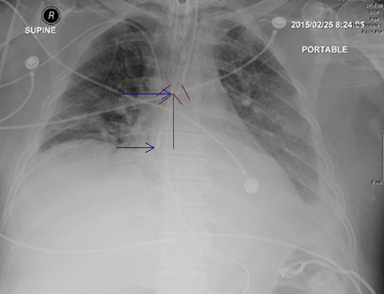
. We told the nurse to check by air bolus while we await a second xray to be completed and read. Chest X-ray demonstrating the Dobhoff tube penetrating the right lung base via the right mainstem bronchus red arrow. Normally the DHT tip should be placed in the 2 nd to 3 rd portion of the duodenum and would create a C-shaped tracing on the X-ray.
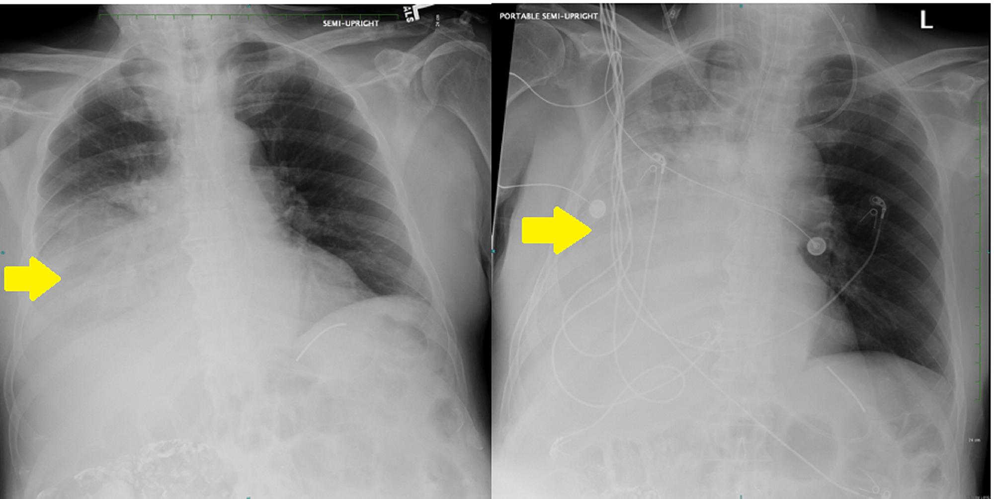
When tube is connected to low intermittent suction there should be return of gastric contents If there isnt confirm placement with xray KUB zWith dobhoff tubes should always confirm placement as no suction will be applied. As seen in Figure 1 the tip of the Dobhoff tube is in the left mainstem bronchus. A review of the x-ray showed that the feeding tube was in the main bronchus.
Just so how do you check the placement of a Dobhoff tube. Department sought to change the Dobhoff NG tube insertion practice to reduce the number of tube placements within the airway. Most tubes are visible on a chest x-ray without a guide wire.
The feeding tube has a weighted metal tip and a guide wire for insertion. The wire was left in. The practice change consisted of implementing the two-step x-ray process which entails the clinician inserting the Dobhoff NG tube to 35 centimeters cm which is at the anatomical position.
A DHT was inserted after intubation for feeding purposes. With CORTRAK 2 you can. An x-ray can ensure that the Dobhoff tube has been placed correctly.
Auscultation was performed in all 78. If we had continued. Tube crosses the diaphragm in the midline.
Thus DHT insertion requires radiologist confirmation of correct placement with chest x-ray CXR increasing clinical delays. Dobhoff tubes come with a radiopaque stripe making them easily visible in. Acute hypoxemic respiratory failure ensued.
Tracheobronchial insertion of DHTs presents a significant risk for pulmonary complications. The tip sits below the diaphragm. It is also important to note that feeding tube insertions causing pulmonary complications are not always related to tracheobronchial insertion.
Most however are placed in the stomach. Therefore bilateral chest tubes were placed. The tube is inserted by the use of a guide wire called the stylet see image1 which removed after the tube correct placement is confirmed.
An electromagnetic stylet provides real-time location information on the tube tip placement within a patients anatomy. In order to prepare a patient for the insertion of a Dobhoff tube the esophagus and nasal cavity are numbed and the patient if conscious may be given a mild sedative. There is often a discussion on whether an OGNG tube should be removed before laryngoscopy and intubation.
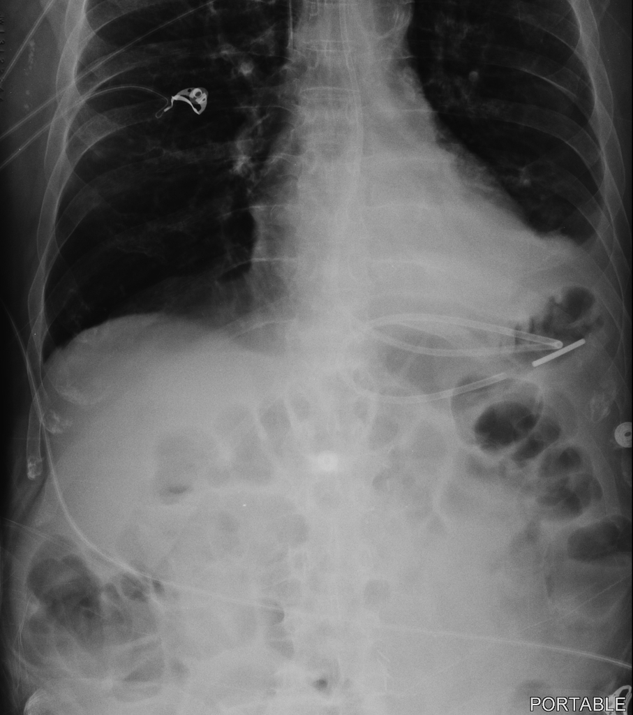
However in our patient who had history of Roux-en-Y the DHT bypassed the duodenum and. An abdominal X-ray was obtained to confirm placement of the DHT Figure 1. ZWith NG tubes placement should be obvious.
Tube bisects the carina. Nasopharynx with 2 Lidocaine jel ly. Dobhoff tubes DHT are narrow-bore flexible devices that deliver enteral nutrition for critically ill patients.
Tip of feeding tube should be in 2 nd or 3 rd portion of duodenum. Of a cough reflex during tube placement is an indication that the tube is being placed in the bronchus rather than the gastrointestinal tract and upon immediate withdrawal of the tube chest x-ray should confirm the absence of pneumothorax. Due to the limitations of bedside techniques in confirming Dobhoff tube placement x-ray remains the gold standard in confirmatory testing 1910.
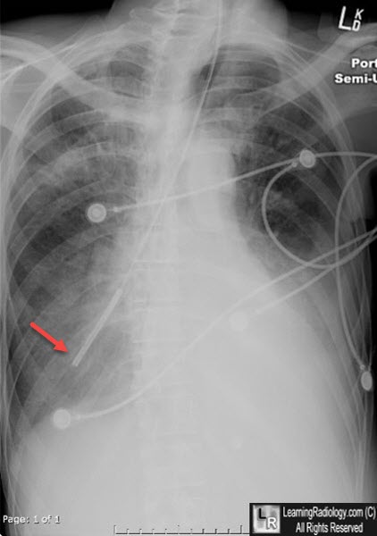
Dobhoff tube is a special type of nasogastric tube NGT which is a small-bore and flexible so it is more comfortable for the patient than the usual NGT. Follow-up abdominal x-ray revealed displacement of the Dobhoff tube in the left pleural space. Of 78 nasoenteral intubations in 46 patients using a Dobbhoff Biosearch Medical Products weighted enteral feeding tube gastric aspirates were evaluated in 28.
Tube descends the thorax in the midline. Once the tube is in the proper place Gastroview contrast is injected into the tube and an image is taken to document the placement. Placement of the flexible nasoenteric feeding tubes in critically ill patients is very difficult.
The x-ray was read and placement confirmed. On-screen visualization provides immediate feedback on tube placement which can reduce or eliminate the need for x-ray confirmation. It was a 12 french by the way.
Data was collected at initial placement prior to x-ray confirmation. Confirmed placement af-ter reading the x-ray. The Dobhoff tube was introduced in the mid-1970s by surgeons Robert Dobbie and Jim.
Steps for NG Feeding Tube Placement in an Awake Patient. The patient was found dead. We had a situation where a dubhoff did not show up anywhere on the x-ray.
We followed the two-step bedside approach that was first described in 19891 First we advanced the tube to 30 centimeters and took a chest x-ray. The patient experi-enced respiratory dis-tress. Tube feedings were begun.
At our facility we x-ray all feeding tubes for placement verification. Two-step approach with a portable chest x-ray to assess accurate and safe placement. You need to be confident that you can see the tip.
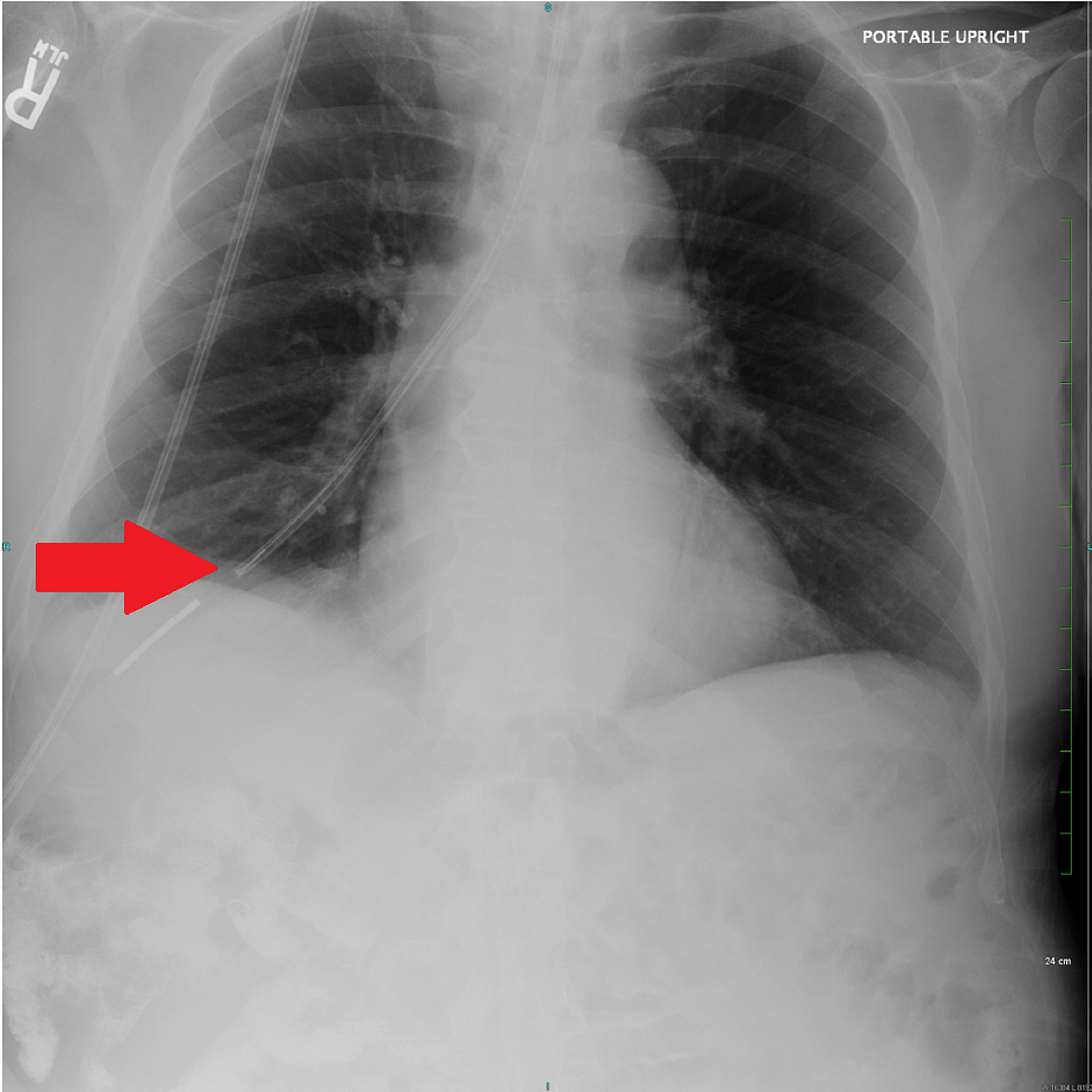
A chest X-ray performed shortly after the tube placement demonstrated that the tip of the Dobhoff tube was within the right lung base following the course of the right mainstem bronchus Figure 1. After insertion correct placement in the body is usually checked with the help of X-rays or fluoroscopy. Immediately identify misplaced tubes.
Measure tube from tip of nose to subxyphoidprocess about 3035cm in most patients Step 2. Place tube through nares and ask patient to swallow as you pass the tube. The nurse is sure of gastric placement.
Feeding tube with guidewire brown tip that is 120cm preferred over blue tip dobhoff tube Lubricant 60 ml syringe. A Dobhoff tube was placed by a house physi-cian. A nasal bandage is used to secure the tube.
After removal of the tube a follow-up chest x-ray revealed iatrogenic bilateral pneumothoraces. Most tube positions are checked by assessing pH of tube aspirate. This short simple video show such a tube in plac.
What can go wrong. They will then insert the Dobhoff tube through the nose into the stomach and into the duodenum. Patients are usually positioned on the right side while the tube is put into the nose.
Abdominal X Ray After Fluoroscopic Guided Dobhoff Tube Placement Small Download Scientific Diagram

Abdominal X Ray Revealing The Dobhoff Tube Traversing The Left Main Download Scientific Diagram
Cureus Hemothorax Following Traumatic Dobhoff Tube Insertion

Iatrogenic Bronchopleural Fistula From A Dobhoff Tube Radiology Case Radiopaedia Org
Cureus Hemothorax Following Traumatic Dobhoff Tube Insertion
Learningradiology Dobhoff Dobbhoff Tube Malplaced Rll
Dobhoff Nasogastric Tube Tube Radiology Case Radiopaedia Org

Icu Chest Radiography Lines Ng Dobhoff Etc Youtube
0 comments
Post a Comment